Nik Noor Ashikin Nik Ab Razak1*, Azhar Abdul Rahman1, Sivamany Kandaiya1, Iskandar Shahrim Mustafa1, Nor Zakiah Yahaya2, Amer Al-Jarrah Mahmoud1, Ramzun Maizan1
1School of Physics, Universiti Sains Malaysia, 18000, Pulau Pinang, Malaysia.
2School of Distance Education, Universiti Sains Malaysia, 18000, Pulau Pinang, Malaysia.
DOI : http://dx.doi.org/10.13005/msri/120101
Article Publishing History
Article Received on : 22 Jan 2015
Article Accepted on : 12 Feb 2015
Article Published : 26 Feb 2015
Plagiarism Check: Yes
Article Metrics
ABSTRACT:
Polymer gel dosimeter is a radiation sensitive chemical dosimeter that can measure 3 D dose distribution with high resolution. Due to the increasing complexity of radiotherapy treatment planning and delivery, accurate experimental radiation dosimetry plays an important role in the implementation and quality assurance of new treatment techniques. A polymer gel dosimeter must possess several important characteristics of a dosimeter to be able to measure absorbed dose precisely. two important dosimetric properties of a dosimeter were determined in this study; accuracy and precision. The MAGAT gels were made of 5% gelatin, 6% methacrylic acid and 10 mM tetrakis-hydroxy-methyl-phosphonium chloride (THPC). The irradiation of MAGAT gel was performed by 6-MV photon beam at a dose range 1 to 10 Gy and was imaged by 1.5 T Magnetic Resonance Imaging (MRI). The dose response of MAGAT gel dosimeter was obtained from spin-spin relaxation rate (R2) of MRI signal. The accuracy of MAGAT gel dosimeter has a range within 4% for doses greater than and equal to 3 Gy. The reproducibility of the MAGAT gel dosimeter at one irradiation was less than 1% whilst the long term reproducibility was within 3% over the five month period. For temporal stability, the dose sensitivity of MAGAT gel dosimeter irradiate at 1 to 11 days post-manufacturing decreased over time. While the dose sensitivity imaged at 1 to 9 days post-irradiation increased up to 4 days post-irradiation and subsequently starts decreasing after 4 days till 9 days. From the study of two dosimetric properties, MAGAT gel dosimeter shows a great dose response with a superior dose response. Thus the MAGAT gel dosimeter can be apply as a 3 D radiotherapy dosimeter.
KEYWORDS:
MAGAT gel dosimeter; spin-spin relaxation rate (R2); dose response; MRI imaging
Copy the following to cite this article:
Razak N. N. A. N. A, Rahman A. A, Kandaiya S, Mustafa I. S, Yahaya N. Z, Mahmoud A. A, Maizan R. Accuracy and Precision of Magat Gel As a Dosimeter. Mat.Sci.Res.India;12(1)
|
Copy the following to cite this URL:
Razak N. N. A. N. A, Rahman A. A, Kandaiya S, Mustafa I. S, Yahaya N. Z, Mahmoud A. A, Maizan R. Accuracy and Precision of Magat Gel As a Dosimeter. Mat.Sci.Res.India;12(1). Available from: http://www.materialsciencejournal.org/?p=1506
|
Introduction
The radiation therapy has been discovered early 1900’s for the treatment of malignant disease using the x-ray radiation. The main principal of radiation therapy is to expose the affected tissue area of a sufficient energy to damage the cells of affected area while sparing the surrounding healthy tissues. The cancerous tissue that been exposed by sufficient levels of radiation, causes a process of irreversible cell damage which preventing the cancerous cells from further growth and metastasis. A potential 3 D dosimeter is required to replace the current radiotherapy dosimetry due to emerging of dynamic radiotherapy applications such as intensity modulated radiotherapy and stereotactic radio surgery. In radiotherapy treatment, the primary goal is to deliver maximum dose to the tumour within the tissue while minimising the dose at surrounding healthy tissue. These conditions may be achieved by tailoring multi-segmented beams to the target volume in which results in reduced absorbed dose of normal healthy tissue thus increased the treatment outcomes. Consequently, this type of treatment required a complex 3D treatment volume in order to verify the complex dose distributions thus a 3D dosimetry is needed. Current radiotherapy dosimetry such as ion chambers, radio graphic film and TLD only offers 1D and 2D dose distributions thus it is difficult to measure the dose over complex 3D treatment volumes.
Radiotherapy gel dosimeter is a chemical dosimeter fabricated from radiation sensitive chemicals in an aqueous gelatin network. Upon irradiation, free radicals from radiolysis process were formed in the gel induce polymerization where amount of polymerization increase and related to absorbed dose (Maryanski et al. 1993 ). Polymer gel dosimetry is one of the gel dosimetry techniques that can be used to verify the spatial dose distribution in complex radiotherapy. Therefore, there exists a need of 3 D dosimeter that has an ideal characterisation of a dosimeter to be used in radiotherapy application.
Methodology
Fabrication of Gel Dosimeter
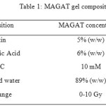
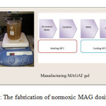
The gel formulation consisted of methacrylic acid (Acros, Organics), gelatin (250 bloom, Bovine) (Sigma Aldrich), de-ionised water, ascorbic acid (Sigma Aldrich) and THPC (Sigma Aldrich). The MAGAT polymer gels were manufactured under normal atmospheric conditions. The gelatin was mixed with de-ionised water in a mixing vessel and was continuously stirred at approximately 48˚C until the gel was completely dissolved and a clear solution was obtained. The solution was cooled to 40˚C then the methacrylic acid monomer was added and continuously stirred until the monomer was completely dissolved. For manufacturing the normoxic polymer gel, an anti-oxidant was finally added to minimise the oxygen exposed to the solution (De Deene et al. 2002a). The fabrication procedures of gel solution are shown as Figure 1. The MAG gels were then poured into 4 ml tissue equivalent polystyrene cuvettes of inner dimensions 1 cm x 1 cm x 4.5 cm ( width x length x height ) with the top sealed by a parafilm tape. Finally they were wrapped in aluminium foils to avoid any preliminary polymerisation from the ambient light. The sample vials were stored at 3˚C before irradiation. In this study, the concentrations of MAGAT gel used are 5% Gel, 6% MAA and 10 mM THPC as shown in Table 1. These concentrations were obtained from our previous study where optimisation of gelatin, MAA and THPC has been done prior to obtain the best sensitivity and dose response of MAGAT gel dosimeter.
Irradiation of gel dosimeter
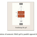
Irradiations were performed using a 6 MV photon beam by a linear accelerator (Primus LINAC, Siemens), with a field size of 10 cm x 10 cmat the isocentre and at 100 cm source axis distance (SAD). The dose rate was 3 Gy min-1. The samples were irradiated from 0 to 10 Gy by parallel opposed beams so that the gels received a uniform dose at 5 cm depth. One sample of each batch is left unirradiated for background measurement. Solid water phantom slabs were placed above and below the Perspex cuvette holder and the samples were placed at the midregion of the phantom as shown in Figure 2. The irradiation were done a day post-manufacturing whereas for long-term reproducibility study, the irradiation was done over a five month period with three batches of MAGAT polymer gel dosimeter were manufactured at different months. Each of the batch was irradiated on 26 Sept 2012, 16 Feb 2013 and 23 Feb 2013. While for post-manufacturing study, the irradiations were done at 1, 4, 8, 11 days post-manufacturing.
MRI Imaging Technique
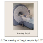
All the sample vials were inserted in a dedicated styrofoam holder and placed in a MRI Signa HDxt 1.5 T whole body scanner using a head coil as shown in Figure 3. All of the samples were imaged a day post-irradiation whereas for post-irradiation study, the MAGAT gel dosimeter were imaged at 1, 2, 4, 7 and 9 days post-irradiation. The imaging sequence applied was a single spin-echo sequence with time echoes of TE1=20 ms and TE2= 300 ms and a relaxation time (TR) of 3500 ms. The other scanning parameters used such as NEX = 3, slice thickness = 5 mm, slice spacing = 0 mm, FOV = 22 mm and flip angle = 90˚. The T2 dicom images were transferred to a personal computer and analyzed using MATLAB 7.1 (Math Works, Inc.) software. From the time series of T2-weighted images (TE=20 ms, and TE=300 ms), R2-maps were calculated from these images for each sequence pixel by pixel basis using pixel signal intensities and applying the two-point method (De Deene and Baldock 2002 b).

The two-point method; where S1, S2 is the measured MR signal intensity at a given echo time, TE and R2 is the transverse relaxation rate. R2 maps can be converted to dose maps using a linear dose response equation that has been reported by several independent investigators (Maryanski et al. 1996a; Ibbott et al. 1997 ; Oldham et al. 1998 ).
R2 = αD + Ro
Linear dose response equation: where α is the slope of the dose–R2 curve, Ro is R2 background and R2 is R2 value of the irradiated gel.
Result and Discussion
The Accuracy of MAGAT Gel Dosimeter
For 3D gel dosimetry, the accuracy and the precision are important factors. The absolute accuracy for basic dosimetry in radiotherapy has to be in the range of 2-3% (Williams and Thwaites). As suggested by The International Commission on Radiation Units (ICRU), the accuracy of absorbed dose has to be within 5 % of the true dose. An accurate measurement is the main requirement of a dosimeter for variety applications of radiation therapy treatments. The accuracy of polymer gel dosimeter is defined as the proximity or the difference between the absorbed dose of gel using the calibrated curve, compared to ‘true value ‘of the measured dose (MacDougall, Pitchford, and Smith 2002). The deviation for MAGAT gel composed of 6% MAA and 5% gelatin had a range within 4% for doses greater than and equal to 3 Gy as shown in Table 2. It is recommended that the gel be irradiated in this dose range in radiotherapy to have the accuracy within 5%.
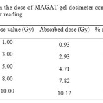 |
Table2: The % deviation in the dose of MAGAT gel dosimeter compared to true dose obtained from the ionisation chamber reading
Click here to View table
|
The Reproducibility of MAGAT Gel Dosimeter
The Reproducibility of MAGAT Gel Dosimeter in one Irradiation
The reproducibility of MAGAT gel dosimeter is expressed as standard deviation in %. Table 3 shows the % variation for reproducibility of MAGAT gel dosimeter at one irradiation was less than 1 %. The small variation in standard deviation indicates the stability of the gel as well as the consistent output of the LINAC.
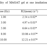 |
Table3: The reproducibility of MAGAT gel at one irradiation represented by % standard deviation
Click here to View table
|
The long-Term Reproducibility in Terms of MAGAT Gel Dosimeter Fabrication
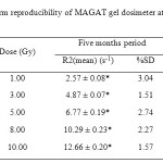
The long term reproducibility of MAGAT gel dosimeter over a five month period is shown in Table 4 with the % standard deviation was within 3%. Although the gel dosimeter is vulnerable to variations of different parameters during the process of gel fabrication it shows a relatively stable response when fabricated at different months. Thus MAGAT polymer gel maybe less sensitive to slight changes in the gel formulation.
Temporal Stability of MAGAT Gel Dosimeter
Post-Manufacturing MAGAT Gel Dosimeter
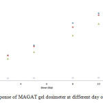
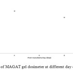
Figure 4 shows the R2-dose response of MAGAT gel dosimeter when irradiated in a time period of 1 to 11 days post-manufacturing. The response trend remains unchanged even though the irradiations were done on different days of post-manufacturing. However, the dose sensitivity of the gel decreased over time as shown in Figure 5. In chemical compounds, the polymerization reaction occurs via a variety of reaction mechanisms that vary in complexity due to the functional group presence in reacting compounds. For MAGAT gel dosimeter, after a week of irradiation the gel shows no response due to the insufficient number of active acrylic monomers to construct the polymer chains through a chemical reaction. The rate of this reaction depends on the intrinsic reactivity of the monomer to radical addition that is influenced by the stability of the monomer towards the addition of a free radical and the stability of the monomer radical. The more stable the radical that is formed from the monomer, the lower the polymerization rate. For the best response, the gel should be irradiated within 24-h post-manufacturing.
Post-Irradiation MAGAT Gel Dosimeter
There are two types of temporal instabilities of the dose response after the polymer gel dosimeter was irradiated. The first instability occurred after the first few hours after irradiation where the polymerization reaction continues, resulting in an increase in the slope of the dose–R2 response. This prolonged reaction is ascribed to the presence of the long-living macroradicals (Baldock, Murry, and Kron 1999). The second instability is when the polymerization process may continue up to a month after the irradiation which is caused by the ongoing gelation of the gelatine, and alters the interception of the dose–response curve (De Deene et al. 2000; Lepage et al. 2001d).
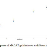
Figure 6 shows the time dependence for MAGAT gel dosimeter via monitoring R2-dose response which changes after being scanned over time period of 1 to 9 days post-irradiation with the common pattern of the response remaining relatively unchanged. Figure 7 shows R2-dose sensitivity of MAGAT gel dosimeter made after being scanned over a time period of 1 to 9 days post-irradiation. The R2-dose sensitivity increased up to 4 days post-irradiation and subsequently starts decreasing after 4 days till 9 days. The temporal stability of the dose response is highly dependent on the type of gel dosimeter. From De Deene study on nPAG gel dosimeter the slopes of nPAG gel dosimeters increase with post-irradiation time (De Deene and Baldock 2002 b) while the slope of the MAGAT gel dosimeter from this study decreases with time. In relative terms, the MAGAT gel dosimeter is more stable compared to nPAG gel dosimeters. It is important to fix the post-irradiation day for imaging. The main factor of an increase in R2-dose sensitivity post-irradiation is due to the continuation of gelation process by gelatine molecules via polymerization reaction caused by the existence of active radicals. It is still unclear on what mechanisms gave a drop in dose sensitivity in the gel. Thus the gel should be imaged after 24 h post-irradiation to avoid continuation of gelling process.
Conclusion
A potential 3 D dosimeter is required to replace the current radiotherapy dosimetry due to emerging of dynamic radiotherapy applications such as intensity modulated radiotherapy and stereotactic radio surgery. A considerable amount of characterisation of normoxic polymer gel dosimeter is required before full implementation of polymer gel dosimetry into the clinical radiotherapy environment is possible. Therefore, it is believed through the findings of this paper provide an important contribution towards the successful implementation of polymer gel dosimetry to clinical radiotherapy.
Acknowledgement
We are indebted to Universiti Sains Malaysia for providing us a short term grant. We would like to thank to Mount Miriam Cancer Hospital, Penang for giving us the opportunity to use the linear accelerator unit and Institut Perubatan dan Pergigian Termaju (Advanced Medical & Dental Institute), Penang for the use of the Magnetic Resonance Imaging unit.
References
- Baldock, C., P. Murry, and T. Kron. 1999. Uncertainty analysis in polymer gel dosimetry. Phys. Med. Biol. 44:N243-N246.
CrossRef
- Baldock, C., R.P. Burford, N. Billingham, G.S. Wagner, S. Patval, R.D. Badawi, and S.F. Keevil. 1998. Experimental procedure for the manufacture and calibration of polyacrylamide gel (PAG) for magnetic resonance imaging (MRI) radiation dosimetry. Phys. Med.Biol. 43:695-702.
CrossRef
- De Deene, Y., and C. Baldock. 2002 b. Optimization of multiple spin-echo sequences for 3D polymer gel dosimetry. Phys. Med.Biol 47:3117-3141.
CrossRef
- De Deene, Y., P. Hanselaer, C. De Wagter, E. Achten, and W. De Neve. 2000. An investigation of the chemical stability of a monomer/polymer gel dosimeter. Phys. Med. Biol 45:859-878.
- CrossRef
- De Deene, Y., C. Hurley, A. Venning, K. Vergote, M. Mather, B..J Healy, and C. Baldock. 2002a. A basic study of some normoxic polymer gel dosimeters Phys. Med. Biol. 47 3441–3463.
CrossRef
- De Deene, Y., N. Reynaert, and C. De Wagter. 2001a. On the accuracy of monomer/polymer gel dosimetry in the proximity of a high-dose-rate 192Ir source. Phys. Med. Biol. 46:2801-2825.
CrossRef
- Doran, S J. 2013. How to perform an optical CT scan: an illustrated guide. Journal of Physics: Conference Series 444 (1):012004.
CrossRef
- Doran, S.J., K.K. Koerkamp, M.A. Bero, P. Jenneson, E.J. Morton, and W.B. Gilboy. 2001 A CCD-based optical CT scanner for high-resolution 3D imaging of radiation dose distributions: equipment specifications, optical simulations and preliminary results Phys. Med. Biol. 46 3191–3213.
CrossRef
- Ibbott, G.S., M.J. Maryanski, P. Eastman, S.D. Holcomb, Y.S. Zhang, R.G. Avison, M. Sanders, and J.C. Gore. 1997 3D visualization and measurement of conformal dose-distributions using MRI of BANG-gel dosimeters Int.J. Radiat. Oncol. Biol. Phys. 38:1097–1103.
CrossRef
- Jirasek, A. 2010. Alternative imaging modalities for polymer gel dosimetry. Journal of Physics: Conference Series 250:012070.
CrossRef
- Jirasek, A.I., C. Duzenli, C. Audet, and J. Eldridge. 2001. Characterization of monomer/crosslinker consumption and polymer formation observed in FT-Raman spectra of irradiated polyacrylamide gels Phys. Med. Biol. 46:151–165.
CrossRef
- Lepage, M., A.K. Whittaker, L. Rintoul, S.A.J. Back, and C. Baldock. 2001d. Modelling of post-irradiation events in polymer gel dosimeters. Phys. Med. Biol. 46:2827-2839.
CrossRef
- MacDougall, N.D., W.G. Pitchford, and M.A. Smith. 2002. A systematic review of the precision and accuracy of dose measurements in photon radiotherapy using polymer and Fricke MRI gel dosimetry. Phys. Med. Biol. 47:R107-R121.
CrossRef
- Maryanski, M.J., J.C. Gore, R.P. Kennan, and R.J. Schulz. 1993 NMR relaxation enhancement in gels polymerized and cross-linked by ionizing radiation: a new approach to 3D dosimetry by MRI Magn. Reson. Imaging 11:253–258.
CrossRef
- Maryanski, M.J., G.S. Ibbott, P. Eastman, R.J. Schulz, and J.C. Gore. 1996a. Radiation therapy dosimetry using resonance imaging of polymer gels Med. Phys. 23 699–705.
CrossRef
- Oldham, M., I.B. Baustert, C. Lord, T.A.R. Smith, M. McJury, M. Leach, A.P. Warrington, and S. Webb. 1998 An investigation into the dosimetry of a 9 field tomotherapy irradiation using BANG-gel dosimetry Phys. Med. Biol. 43 1113–1132.
CrossRef
- Williams, J.R. , and D.I. Thwaites. Radiotherapy physics in practice Second edition.
Views: 1,366
 This work is licensed under a Creative Commons Attribution 4.0 International License.
 Material Science Research India An International Peer Reviewed Research Journal

